
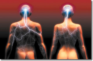
Male and female brains are wired differently. That is not intended metaphorically – they literally have different amounts and/or patterns of axonal connections between a variety of brain regions, as well as differences in the size or number of cells in various regions. This is true in mammals, birds, fish, even insects and correlates with hard-wired, innate differences in behaviour between the sexes across in species across all these phyla. This is as true for humans as for any other species. The behaviours that show the most robust and innate differences between the sexes are involved in mating, reproduction, parental behaviour, territoriality and aggression and it is the brain areas that control these behaviours that are the most obviously sexually dimorphic (showing a difference in size or morphology between the sexes). In mammals, these include areas in the limbic system, including parts of the hypothalamus, amygdala, preoptic area and bed nucleus of the stria terminalis.
How do these differences come about? Sex in mammals is determined by the presence of a specific gene, Sry, on the Y chromosome – this gene sets in motion a cascade of gene expression and biochemical changes which lead to the conversion of the undifferentiated gonads into testes in male embryos. (In female embryos the gonads follow a default differentiation pathway to form ovaries). Testes make testosterone, of course, and testosterone is essential to masculinise the developing embryo, so that it develops male external genitalia and other physical sexual characteristics.
Testosterone is also essential to masculinise the brain, but exactly how it does this is very surprising. This phenomenon has been well studied in rodents but the principles apply across mammalian species. In male mice and rats there is a surge in the production of testosterone shortly after birth, which lasts for a couple of days. This surge is precisely timed with a critical period of brain development , during which it is susceptible to the effects of testosterone. If male rats are castrated at this age (I know, sorry), then their brain will develop in the female pattern and they will not display typical male behaviours in adulthood, even when supplemented with testosterone. Conversely, if female rats are given a single dose of testosterone a day or two after birth, their brains develop a male morphological pattern and they will show male-typical behaviours (mounting other females and decreased receptivity to being mounted). Crucially, the same manipulations carried out a week or two later have no effect on either brain morphology or behaviour.
These effects of testosterone are called “organisational”, for obvious reasons, and are distinguished from the later effects in acutely stimulating male behaviours, which are called “activational”. Now for the surprise. It was also found that a single dose of estrogen, given to postnatal female rats, was just as effective as the testosterone in masculinising their brains – even more effective, in fact! How could this be? How can estrogen have the same effects on the developing brain as testosterone? As it turns out, testosterone is actively converted into estrogen in the brain, through the action of an enzyme, aromatase. This enzyme is specifically enriched in brain regions that show sexual dimorphism. The estrogen then acts through two estrogen receptors and this activity has been shown to be required for the masculinising effects of testosterone. In particular, mutations in aromatase or in the estrogen receptors block the effects of testosterone and result in male animals with female brain morphology and behaviour, despite normal levels of circulating testosterone.
So, why don’t females have masculinised brains? They should have loads of estrogen, shouldn’t they? In fact female rodents have very low levels of circulating estrogen at this early postnatal stage, coinciding with the critical period.
The surprising findings implicating aromatase and estrogen receptors have left a mystery surrounding the role of the androgen receptor – the protein traditionally associated with direct responses to testosterone. Mice with mutations in the gene for the androgen receptor also show feminised behaviours, suggesting it is also important in the process of masculinisation. However, this interpretation is complicated by the fact that these mutants also show testicular atrophy (sorry again) and consequently have very low levels of circulating testosterone. A new study by Nirao Shah and colleagues has now resolved the role of the androgen receptor in controlling sexual behaviour.
By knocking out this gene just in the brain, they managed to get around the requirements for testicular function and so cleanly address the possible functions in the brain. They clearly show that the brains of these conditional mutants are still masculinised, morphologically. These mice also generally show a male pattern of behaviour. However, they do not express all of these behaviours to the same extent as wild-type males. In particular, the frequency of mating behaviours in the presence of an estrus female is reduced, though when they do engage in the behaviour the routine is effectively normal. The mutant males also mark their territory less than wild-types and spend less time fighting with other “intruder” males.
Thus, while the developmental effects of testosterone appear to be fully explained by its conversion to estrogen, the activational effects in adults which are required for the full expression of male behaviours depend at least in part on its direct action through the androgen receptor.
Wu, M., Manoli, D., Fraser, E., Coats, J., Tollkuhn, J., Honda, S., Harada, N., & Shah, N. (2009). Estrogen Masculinizes Neural Pathways and Sex-Specific Behaviors Cell, 139 (1), 61-72 DOI: 10.1016/j.cell.2009.07.036
Scott A. Juntti, Jessica Tollkuhn, Melody V. Wu, Eleanor J. Fraser, Taylor Soderborg, Stella Tan,, & Shin-Ichiro Honda, Nobuhiro Harada, and Nirao M. Shah (2010). The Androgen Receptor Governs the Execution, but Not Programming, of Male Sexual and Territorial Behaviors Neuron, 66, 260-272 : DOI 10.1016/j.neuron.2010.03.024


