Are bigger bits of brains better?
")
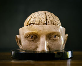
We scoff at the folly of phrenology – the simplistic idea that the size and shape of bumps on the skull could tell you something about a person’s character and psychological attributes. It was all the rage in the Victorian era (the early to mid-1800s) in the UK and the US especially, with practitioners armed with calipers claiming to measure all kinds of personal propensities, from Acquisitiveness and Combativeness to Benevolence and Wonder. The skull bumps were just a proxy, of course – the idea was that they reflected the size and shape of the underlying brain regions, which were what was really associated with various traits. It all seems a bit quaint and simplistic now (apart from the entrenched association with racism), but while we may like to think we have moved on, a lot of modern human neuroscience is founded on the same premises.
- The first premise is that different mental functions or psychological traits can be localised to specific regions of the brain.
- The second is that the size of those regions is correlated with the level of function or trait – usually with the idea that bigger is better.
- And there is a third premise, which also sometimes comes along for the ride, which is captured by the slogan “use it or lose it” – the idea that if some brain area or function it supports is not used, that brain area will get smaller; the converse idea is that if it is used more, it will get bigger.
These ideas are pervasive in the public perception of neuroscience, promoted, in my view, by the way we neuroscientists tell our stories. But this is not just a problem of communication of science – these assumptions are also implicit and typically unexamined in the motivation for and interpretation of many studies in the field.
They weren’t plucked out of the air, of course – there is some underlying truth to all of these ideas, and a relevant evidence base supporting them. For example, lesion studies, patterns of brain activation during various tasks, and the selective effects of stimulation of various bits of the brain all support the partial localisation of all kinds of cognitive functions. However, none of these kinds of evidence suggest that the implicated brain regions carry out these functions by themselves. We now understand that most cognitive functions are mediated by distributed networks, not by isolated brain regions. There is certainly a high degree of specialisation of function in that extended circuitry and it’s interesting and important to map that out, but I think we’d all hope to have moved beyond naïve “blobology”.
Similarly, while there are plenty of examples where size really does matter for some function or other, in both animals and humans, it is not obvious that equivalences can be drawn across the range of scenarios where this seems to hold. Differences in size and function (and correlations between them) have been studied in many species across evolution, across development, across the lifespan, across individuals, between sexes, across seasons, in pathological states, and in response to experience. Findings from one of these areas are often used to bolster claims or motivate experiments in another, but disparate underlying mechanisms may be at play across these diverse scenarios.
This is especially true in human studies as the only non-invasive way to measure the size of bits of brains in humans is by neuroimaging, which is extremely crude, relative to techniques that can be applied to animal brains. Methods like voxel-based morphometry (VBM) let you gauge the size of different bits of the brain across individuals by warping 3D images of their brains to a common template. You can similarly measure thickness or surface area of bits of the cortex and compare across individuals. But there are all kinds of different parameters at a cellular level that could drive variation in the overall size of bits of the brain, which may be indistinguishable by neuroimaging.
These parameters include numbers of neurons, numbers of astrocytes and oligodendrocytes, cell sizes, the extent of dendritic or axonal arbors, numbers of synapses, myelination, vascularisation, or many others. And each of those parameters is itself determined by multiple distinct processes, such as proliferation, differentiation, migration, survival, growth, pruning, and on and on. Very different processes and parameters will be at play in different situations.
To take a few examples, a difference in size of some region over evolution may be caused by changes in proliferation and neurogenesis, which could in turn have various underlying causes, such as direct effects on cell cycle genes or indirect effects caused by differences in afferent connectivity and the supply of growth factors; changes with aging may track death of neurons or glial cells, loss of myelin or other degenerative processes; while differences across individuals could be due to variation in neuronal number or in things like the elaboration of neurites and synapses – the same number of neurons, but with bushier connections. All of these may manifest as a difference in size of some region by neuroimaging, but they’re not clearly meaningfully related to each other.
The causes of size differences are thus quite diverse. What about the consequences? Is bigger necessarily better? There certainly are cases where it is, but again, it’s important not to conflate these or compare apples with oranges.
When it comes to the whole brain, size certainly matters. Brain size correlates well with intelligence across species, with bigger-brained species mostly being more intelligent, and especially a consistent trend of increasing size in the primate lineage leading to humans. And if we look across humans, whole brain size also correlates with intelligence (measured in any of a variety of ways), with correlation coefficients in the range of r=0.2–0.3.

There is an interesting exception to that, however. Average male brain size in humans is about 10% larger than average female brain size, but there is no difference in mean I.Q. between the sexes. This suggests some other sex difference (in functional organisation, perhaps) counteracts the size effect on I.Q., which otherwise holds within each sex. Even for the whole brain, size clearly isn’t everything, a principle which holds true across species too.
Whole brain size is also obviously the grossest neuroanatomical measure you can get. What about smaller bits of the brain? Does the size of any of them correlate with function or variation in traits? If you’ve been reading the human neuroimaging literature for the last couple of decades or the popular media describing it you would certainly be convinced that many examples exist of such a relationship.
I would guess there are thousands of published papers reporting such an association. But I can’t think of any that have robustly replicated and there is good evidence to suggest that most such reports are spurious findings.
A very recent paper by Marek and colleagues provides compelling evidence that most such studies have been statistically underpowered by a couple orders of magnitude. Where sample sizes have typically been in the tens or low hundreds, this paper shows that you need to get to samples in the range of 10,000 people before you can detect robust associations between neuroimaging measures and behavioural phenotypes. Below that, sample variance leads to wildly fluctuating results, producing many false positive “findings” and hugely inflated effect sizes. The pernicious effect of publication bias massively exacerbates the problem in the literature. (The similarities to the candidate gene association literature are not coincidental).
Moreover, even for the positive associations that were found in this study, they were with extremely broad psychological phenotypes – general cognitive ability and psychopathology – and the anatomical measures associated with them were highly distributed across the whole brain. What about more specific associations between the sizes of bits of the brain and more specific traits or functions? Is there any reason to think they should exist?
Clearly, we could reasonably expect that the variation that we observe in psychological traits is caused by variation in some parameters in the brain, but should we expect them to be manifested at the macroscopic level probed by neuroimaging? Or are we just taking that approach in humans because no other methodology is available? Are we doing it just because it’s the only thing we can do, not because there is a principled reason to expect such associations to exist?
If the entirety of the prior literature reporting such associations is now in question, is there any other evidence base for the hypothesis that the size of specific brain regions is associated with degree of function? Well, yes, though most of the evidence that springs to my mind is somewhat indirect, for example from pathological situations, from studies looking over evolutionary timescales, or from seasonal changes in some animals that may be quite unusual.
In many neuropathological conditions, or even in normal aging, the degree of loss of grey or white matter often correlates with the severity of neurological or psychological symptoms. However, as with lesion studies, there is a limit to how far this kind of relationship can be extrapolated – greater loss of neural tissue in pathological situations is certainly worse, but it’s not clear that having more in a given area in a healthy situation (i.e., across individuals in the healthy population) would necessarily be better for some specific function.
In evolution, there are lots of examples of differences in size of brain regions that correlate with behavioural adaptations across species. For example, in mammals that have evolved to use various senses to different degrees, those senses that are used more have a larger representation in the cerebral cortex. Different species may literally need more neural real estate to effectively process the richer signals coming in through the visual or auditory or olfactory domains. There’s no point having bigger eyes with more photoreceptors of different kinds enabling more sophisticated visual processing if the brain can’t handle that information.

It’s interesting to note that the underlying mechanisms involve a dynamic communication between different parts of the nervous system, from the sensory periphery through all the relays to the higher perceptual centres. The size of any given brain region is not determined solely by a genetic program that runs in isolation in those cells – it is also affected by proliferation signals and neurotrophic factors released by incoming nerve fibres from other regions in the circuit. Thus, over both development in individuals and the evolution of species, the size of functionally coupled regions is tightly coordinated. (Indeed, cortical thickness within individuals is correlated between regions that make up extended functional circuits).
An even more dynamic relationship between size and function is observed in some animals with seasonal changes in the size of some brain region. For example, in some male songbirds, the high vocal centre or “song nucleus” grows in response to changes in day length and hormonal signals, giving them more neural resources for the mating season, when their singing needs to be on point. Similarly, some mammals and birds show changes in the size of the hippocampus across seasons, which may reflect different behavioural demands on navigational abilities across the seasons. Such changes involve high levels of neural turnover and proliferation of new neurons, tightly regulated by a variety of signaling mechanisms.
These seasonal changes thus represent good examples of all three premises: particular behavioural functions in these species do rely (especially though not exclusively) on these specific brain areas; the size of these areas does seem to reflect degree of functional capability; and when they are not needed at full capacity they shrink (presumably due to an otherwise unnecessarily high metabolic cost of maintaining them).
Though humans don’t show such seasonal changes, there are numerous reports of differences in brain region size that are supposedly driven by differences in experience and associated with the level of some cognitive function. The most famous of these reports involve London taxi drivers, who were found to have larger hippocampi (specifically the posterior portion) than non-taxi-driving subjects. The interpretation was that this difference was driven by their intense training on the geography of London streets (“the knowledge”), with the implication at least that this increase in size improved their navigational abilities.
[Note added: See also a subsequent paper finding a change in hippocampal size following training:
Acquiring “the Knowledge” of London's Layout Drives Structural Brain Changes]
These studies are very well known and effectively taken as lore, both in the field and by the general public. (I have had taxi drivers themselves tell me about these findings). Without dwelling on it, I think the design of the original studies would not stand up well to current expectations, especially in terms of sample sizes, which were in the tens (not the tens of thousands). Moreover, larger studies have not observed a relationship in the general population between size of the hippocampus (or bits of it) and navigational abilities.
Again, the rationale for the hypothesis that bits of the brain should grow with use is rather vague. Are we expecting new neurons to be produced? (This might be the case in the hippocampus though even there the question of whether adult neurogenesis actually occurs in humans is controversial). Or would it be driven by growth of dendrites and synapses? Or maybe very active regions get swole by attracting a bigger supply of blood vessels?
But what does “very active” even mean here? This idea harkens back to the myth that we only use 10% of our brains. Really we’re using all of our brains, all the time, and even when we’re not obviously actively “doing something” with some brain region, there’s still loads of background neural activity there.
Are the hippocampi of taxi drivers really more active than those of other folks? I don’t just mean that they are using them more to navigate – I mean that if you measured the actual neural activity in those structures over some period of time, it would be on average higher (to an extent that might plausibly drive a cellular physiological response and growth of the whole area). That might be true but I don’t think it’s been shown.
Or are they in some way differently active? Maybe there’s more high-frequency firing or more synchronised firing or variation in some other global parameter without a change in overall number of spikes. Perhaps cells could monitor and respond to such parameters. There is an extensive body of literature in animals showing that both levels and patterns of neural activity do feed back onto gene expression in ways that are important for both activity-dependent development and plasticity. As far as I know, however, these have not been linked to macroscopic growth of whole areas but rather to highly cell-type-specific microscopic changes in connectivity.
More generally, it is true that synaptic plasticity and learning, especially the formation of long-term memories, involves the physical growth of new synaptic connections. However, this is not one-way: synaptic plasticity equally involves the weakening and pruning of connections under other conditions. Moreover, absolute increases in overall numbers or strength of synapses are actively counteracted by homeostatic processes that reduce overall synaptic connectivity (especially during sleep), maintaining relative changes in strength, while renormalizing the responsiveness of the network to new experience.
In short, brain is not like muscle. Bits of brain don’t just grow with experience – they mainly change by reorganising their internal connectivity. This is just as well because if the brain did continue to grow with use, all of our brains would be busting out of our skulls. I don’t mean to be too sarcastic (just the right amount), but I’ve been going around seeing things like crazy – really intensely using my visual system for many years now – without causing massive growth of my visual cortex.
So, where does all this leave us? Let’s reconsider our three premises:
Can we localise functions to specific brain areas? We can certainly implicate areas as being involved more in or required more for some functions than others, without falling into the trap of thinking that they are somehow sufficient for those functions. (You can say the same for genes).
Is bigger better? In many scenarios – evolutionary, developmental, pathological – the answer is clearly yes. I’m sure that readers will think of many other examples that fit with this general idea. But to return to where we started, with phrenology, those observations do not necessarily imply that variation in size of bits of brain should contribute to variation across the normal range of behavioural traits or cognitive functions within humans.
Should we expect variation in extraversion or impulsivity or murderousness or spatial reasoning or working memory or creativity be associated with the macroscopic size of specific brain regions, as measurable by neuroimaging? Maybe. It’s not an entirely daft idea, it’s just really crude and simplistic, in my opinion. An alternative hypothesis that I find more likely is that variation in those kinds of complex psychological traits and cognitive functions will be due to idiosyncratic combinations of distributed and likely subtle effects on all kinds of cellular and biochemical parameters affecting the function and computational operations of highly extended circuitry across the brain. Complex traits are called that for a reason.
And finally, do bits of brain grow with use in a way that mediates greater function? The motivation for this hypothesis is vague and weak, in my opinion, and though it is taken as lore, the evidence for this kind of effect in humans is on far shakier ground than many people both in the field and in the general public appreciate.
Generally speaking, while our technology has improved, we won’t make progress in understanding the complex relationships between human brain structure and function and our individual psychological traits armed only with Victorian-era theories.

Comments
Post a Comment