Do you see what I see?
 An enduring question in philosophy and
neuroscience is whether any individual’s subjective perceptual experiences are
the same as those of other people. Do you experience a particular shade of red
the same way I do? We can both point to something in the outside world and
agree that it’s red, based on our both having learned that things causing that
perceptual experience are called “red”. But whether the internal subjective
experience of that percept is really the same is almost impossible to tell.
An enduring question in philosophy and
neuroscience is whether any individual’s subjective perceptual experiences are
the same as those of other people. Do you experience a particular shade of red
the same way I do? We can both point to something in the outside world and
agree that it’s red, based on our both having learned that things causing that
perceptual experience are called “red”. But whether the internal subjective
experience of that percept is really the same is almost impossible to tell.
There are some exceptions, of course, where
there are clear differences between people’s perceptions. Colour blindness is
the most obvious, where individuals clearly do not experience visual stimuli in
the same way as non-colourblind people. This can be contrasted with the
experience of people who are tetrachromatic – who can distinguish between a
greater number of colours, due to expression of a fourth opsin gene variant. Conditions
like face blindness and dyslexia may involve difficulties in higher-order
processing of specific categories of visual inputs. And synaesthesia provides a
striking example of a difference in subjective perceptual experience, where
certain stimuli (such as sounds, musical notes, graphemes, odours or many other
inducers) are accompanied by an extra visual percept (or percept or association
in some other modality).
But what about more general experience? Do
people without such striking conditions exhibit stable individual differences
in how they see things? Geraint Rees and colleagues have done some fascinating
work to show that they do and also linked such differences in subjective
experience to differences in the size of the visual cortex.

They have used a number of visual illusions
as tools to quantify individuals’ subjective experience. One of these is the
well-known Ebbinghaus illusion, where a circle seems to differ in size when
surrounded by either bigger or smaller circles. (Even though you know it’s the
same size it’s almost impossible to see it that way). Rees and colleagues
assayed how susceptible people were to this illusion by asking them to match
the perceived size of the internal circle with one of a set of circles that
really did vary in size.

They then used functional neuroimaging to
map the spatial extent of the primary visual cortex (area V1) in these people.
This is the first region of the cerebral cortex that receives visual
information. This information is conveyed by direct connections from the dorsal lateral geniculate nucleus (dLGN), the visual part of the thalamus, which
itself receives inputs from the retina. The important thing about the
projections of nerve fibres from the retina to the dLGN and from there to area
V1, is that they form an orderly map. Neurons that are next to each other in
the retina project to target neurons that are next to each other in the dLGN
and so on, up to V1. Since the visual world is itself mapped by the lens across
the two-dimensional surface of the retina, this means that it is also mapped
across the surface of V1.
Rees and colleagues took advantage of this
feature to map the extent of V1 using functional magnetic resonance imaging
(fMRI). By moving a stimulus across the visual field one gets an orderly
response of neurons from different parts of V1, until a point is reached at
which the responses reverse – this is the start of the second visual area, V2.

Remarkably, the strength of the visual
illusion (and of another one called the Ponzo illusion) experienced by individuals correlated strongly (and negatively) with
the size of their V1. That is, individuals with a smaller V1 experienced the
illusion more strongly – they were the least accurate in judging the true size
of the inner circle. Put another way, their perception of the inner circle was
more affected by the nearby outer circles. This suggests a possible explanation
for this effect.
Neurons in V1 receive inputs from the dLGN
but also engage in lateral interactions with nearby V1 neurons. These integrate
responses from neighbouring visual fields and help sharpen response to areas of
higher contrast, such as edges of objects. If the visual world is projected
across a physically smaller sheet of neurons, then the responses of neurons in
one part may be more affected by neighbouring neurons responding to nearby
visual stimuli (the outer circles in this example). Conversely, a larger V1
could mean that each neuron integrates across a smaller visual field,
increasing visual resolution generally and reducing responsiveness to the
Ebbinghaus illusion.

That makes a pretty neat explanation of
that effect (probably too neat and simple, but a good working hypothesis), but
leads us on to another question. How do differences in the size of V1 come
about? What factors determine the spatial extent of the primary visual cortex
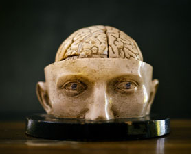
determined? There is considerable variation in this parameter across
individuals, as assessed by neuroimaging or by post mortem cytoarchitecture (as
in the diagram, showing the extent of V1, labelled as 17 and of V2, labelled as
18, in eight individuals). Is the extent of V1 genetically determined or more
dependent on experience?
The heritability of the extent of V1 itself
has not been directly studied, to my knowledge, but the surface area of the
occipital lobe (encompassing V1 and other visual areas) is moderately heritable
(h2 between 0.31 and 0.64 in one study). This is supported by a more recent twin study, which used genetic correlations to parcellate the brain and also found
that the surface area of the entire occipital lobe is heritable, largely
independently of other regions of the brain. (Another reason to expect the
extent of V1 to be heritable is that it is very highly correlated (r=0.63) with
the peak gamma frequency of visual evoked potentials as measured by EEG or MEG.
This electrophysiological parameter is a stable trait and has itself been shown
to be extremely highly heritable (h2=91%!)). Size and shape of cortical areas also vary between inbred mouse strains, demonstrating strong genetic effects on these parameters.
Interestingly, cortical thickness and
cortical surface area are independently heritable. This is consistent with the
radial unit hypothesis of cortical development, which suggests that the surface
area will depend on the number of columns produced while the thickness will
depend on the number of cells per column. These parameters are likely affected
by variation in distinct cellular processes.

What could these processes be? What kinds of
genes might affect the surface area of V1? One class could be involved in early
patterning of the cortical sheet. A kind of competition between molecular gradients from the front and back of the embryonic brain determines the
relative extent of anterior versus more posterior cortical areas. Mutations
affecting these genes in mice can lead to sometimes dramatic increases or
decreases in the extent of V1 (and other areas). A negative correlation that has been observed between
size of V1 and size of prefrontal cortex in humans might be consistent with
such an antagonistic model of cortical patterning. This mechanism establishes
the basic layout of the cortical areas, but is only the first step.
The full emergence of cortical areas
depends on their being innervated by axons from the thalamus. For example,
axons from the dLGN release an unidentified factor that affects cell division
in V1, driving the expansion of this area. The size of the dLGN is thus
ultimately correlated with that of V1. In addition, the maturation of V1,
including the emergence of patterns of gene expression, the local cellular
organisation and even the connectivity with other cortical areas all depend on
it being appropriately innervated. Variation in genes controlling this
innervation could thus indirectly affect V1 size.
Axons from the dLGN are specifically guided
to V1 by molecular cues, though the identity of these cues remains largely
mysterious. For example, my own lab has shown – in studies of a line of mice
with a mutation in an axon guidance factor, Semaphorin-6A – that even if dLGN
axons are initially misrouted to the amygdala, they eventually find their way
specifically to V1 and are even able to evict interloping axons from
somatosensory thalamus, which had invaded this vacant territory. Not all the
misrouted axons make it to V1, however, and many that do not eventually die.
The end result is that the dLGN is smaller than normal and V1 is also smaller. I am not suggesting that this
specific scenario contributes to variation in V1 size in humans
but it illustrates the general point that the number of dLGN axons reaching V1
is another factor that will affect its ultimate size.
Whatever the mechanisms, the studies by
Rees and colleagues clearly demonstrate considerable variation in subjective
visual experience across the population and provide a plausible explanation for
this in a heritable variation in brain structure. So, the short answer to the
question in the title is most probably “No”. (And the long answer is already
way too long, so I’ll stop!)


Fascinating as ever.
ReplyDeleteThere's a potential autism connection here too in that it's widely claimed that people with autism are less susceptible to visual illusions such as the Ebbinghaus. This would suggest an interesting experiment looking at the extent of V1 in autism using Rees' technique.
An important caveat though is that the evidence is pretty inconsistent. The original study by Happe 1996 which everyone cites pretty much involved giving everyone the illusion and asking "can you see it?" Later studies by Ropar and Mitchell included control conditions where the circles really are different - or involved adjusting the circles so they're the same. And they found no group differences - so obviously much less cited! But then again some more recent studies do seem to find differences.
So perhaps as with all things autism, there are some people who are less susceptible and others more so. Which would again be a good reason to look at it in relation to V1 - but in terms of individual differences, which may be more exaggerated than in the non-autistic population.
Thanks Jon. That's very interesting to get a critique of the widely cited Happe paper. You would think more people would have studied that. The technique by Rees and colleagues for quantifying the effect of the illusion seems like it could readily be applied to assess if there is a real difference in people with autism.
DeleteThe recent paper by Pellicano and Burr (http://www.ncbi.nlm.nih.gov/pubmed/22959875) suggests top-down explanations for reduced susceptibility to illusions in terms of weaker Bayesian priors, as opposed to bottom-up explanations, though I know you argued the reverse in reply to it:
(http://www.ncbi.nlm.nih.gov/pubmed/23123383)
I wasn't so much arguing the reverse as pointing out that the reverse would give you very similar predictions - so it's not clear what the Bayesian angle gives you (although it does offer the intriguing possibility that different underlying mechanisms in different individuals could converge on very similar perceptual experiences)
DeletePellicano and Burr's rebuttal I think missed the point because they ended up preferring their "reduced priors" account on the grounds of the neurophysiological implausibility of "reduced neural noise" - which I was thinking of in more abstract terms than actual neurons! And in any case, they could have come to the same conclusion without writing a paper on Bayesian perception!
Planning a blogpost on this at some stage - but trying to get my head around Karl Friston's comment first!!
The classic twin study design can demonstrate if a condition has a strong genetic component but cannot accuratly calculate a 'heritabilty estimate' based on the difference in concordance rates between MZ and DZ twins. The rate of de novo gene mutations caused by reproductive error (egg or sperm)has inflated the 'heritability' of disorders like autism and schizophrenia.
ReplyDeletehttp://www.ncbi.nlm.nih.gov/pubmed/22914163
Paradoxically in Down syndrome which is almost always caused by a random reproductive error and is mot inherited, concordance in MZ twins for Down Syndrome is nearly 100% and concordance in DZ twins is nearly 0% which would make Downs syndrome the most 'heritable' of all the developmental disorders.
You're right that heritability estimates will be increased by de novo mutations, which will always be shared by MZ twins and never by DZ twins. And this is indeed an important factor in SZ and ASD. So I agree that the exact number that emerges from twin studies is less important than indicating something is heritable per se.
DeleteAnd Down syndrome is entirely heritable. That does not mean it's inherited - it means that all of the variance in the population in whether people get Down syndrome or not is entirely explained by genetic variance (in this case, an extra chromosome 21).
All genetic muations including de novo gene mutations are heritable only in that they are derived from a paternal or maternal gene even though the parent does not possess the genetic mutation.
ReplyDeleteCase in point, Klinefelter Syndrome is not 'inherited' and is always caused by a random sperm or egg mutation. 53% of the cases are derived from a paternal sperm muation and 47% are derived from a maternal egg mutation.
Regarding the concepts of environment vs genetic causation, Klinefleter Syndrome is a strongly genetic disorder but is primarily caused by environmental risk factors.
The several studies found that PCB congeners, as measured in blood, produced XY sperm mutations and the frequency of XY sperm mutations increased with increasing levels of PCB congeners. Another study found that that the frequency of XY sperm increased with advancing paternal age
http://www.ncbi.nlm.nih.gov/pmc/articles/PMC3339457/
http://www.ncbi.nlm.nih.gov/pmc/articles/PMC1274351/
Environmnetal risk factors are central in understanding the mechanisms underlying sperm or egg mutations in healthy parents.
You're right that the concept of heritability can be ambiguous, depending on perspective. On the one hand, you can ask: is the proximal cause of a disorder genetic? It may be completely genetic as in Klinefelter or Down syndrome. On the other hand, it may be true that the genetic difference is itself caused by some non-genetic factor, such as an environmental exposure. So, the difference across the population in whether people get the disorder or not could be entirely environmental. (Or it could just be bad luck).
DeleteI had nearly given up on this blog after just having discovered it, as it seemed to have gone quiet for a while there. However, whilst there is some current commentary, and in spite of the fact that I do not hold a degree in neuroscience, nor did I even get past 10th Grade way back in the 60's for that matter, I would like to write one thing.. , I am more and more fascinated by the workings of the brain as time goes on, and despite the degree of academic literature out there on this subject, there is so very little we really still do understand about the brain. I shall be fascinated and always interested at least by this fact alone, until my days are up, mainly due to the combined facts that I, first of all, do have a cavus septum pellucidum which I believe holds far more undiscovered implications than the scientific fraternity care to try to understand, much less to explore in depth! Secondly, my dear husband suffers from the worst kind of RLS (or now more correctly renamed Willis-Ekbom Disease in order hopefully, for the medical fraternity to treat it with the respect it actually deserves).. and has done for years, and has taken medication 24/7 for years to at least try to control the symptoms.
ReplyDeleteAll of the origins of both anomalies of the neuro system still have far more investigative issues to be addressed than anyone in the relevant fields apparently is willing to do at this point in time, unfortunately.
But thanks for the opportunity to pen my thoughts here in this blog.. I can provide quite a bit of history and opinion on both these conditions, but then, I hardly qualify from an academic standpoint! Thanks for listening and thanks for the blog!
Information and articles posted on this site are really useful to the readers. Keep up the good work on sharing your great ideas.
ReplyDelete